Corneal Topography

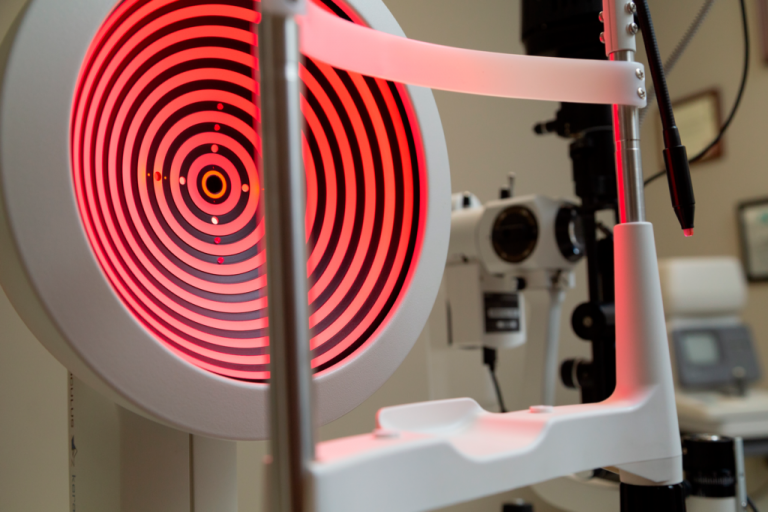
Corneal topography, also known as corneal mapping, is a diagnostic tool that provides three-dimensional images of the cornea.
The cornea is the anterior, clear, central layer of the eye, responsible for about 70% of the refractive power of the eye. Corneal topography provides detailed 3D maps of the shape and curvature of the cornea and allows the detection of corneal diseases and irregular corneal conditions, such as swelling, scarring, abrasions, deformities and irregular astigmatisms.
Corneal topography is also used for the monitoring and treatment of corneal diseases, laser design, eye surgery and even the adaptation of contact lenses.
The examination is contactless and painless and usually takes place within 2-3 minutes.

Types of corneal topography
There are three different types of technologies used for corneal topography:
Placido Disc Topography
Placido disc reflection systems measure curvature, abnormalities, quality of lacrimal film, foreign bodies and other parts of the anterior cornea. Reflection depends to a large extent on the tear film that reflects light and can be either a small or a large focus; small focuses are more accurate as they collect more data points, but large focuses are easier to use and make data collection significantly easier.
Scheimpflug topography and scanning
These two systems provide information about the anterior and posterior corneal surface and are used to detect and manage corneal swelling, which is especially important for contact lens users.
Types of topographical maps
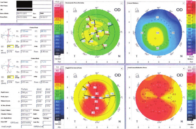
Axial display map
This is the most traditional way to view a topographic image, as it is useful for an overview of corneal strength.
Tangent map view
This type of map provides an accurate measurement of the power and curvature of the cornea and is therefore useful in the placement of contact lenses, especially ortho-k lenses.

Elevation map
This map is used to determine the actual shape of the cornea and is crucial for choosing the best contact lens design for an irregular cornea. This map visualization is especially important when deciding between a permeable gas scleral (GP) or corneal contact lens.
Corneal thickness display map
This map illustration is used for staging of corneal diseases, such as keratoconus, but is mainly used to monitor changes in corneal thickness when wearing contact lenses.
Tear split screen
This map shows the quality of the natural tear layer and also shows whether the quality of tears has been affected by the wear of contact lenses.
Indications:
- Keratoconus
- Laser refractive surgery design (LASIK, PRK)
- Monitoring of the course following refractive laser surgery
- Identification of an appropriate intraocular lens for cataract surgery
- Evaluation and treatment of astigmatism after corneal transplantation
- Assessment of corneal damage such as reeds, corneal scars and Salzmann nodes
- Assessment of the integrity of the anterior angle for glaucoma patients
- Corneal thickness measurement
- Detection of infectious keratitis
- Examination of the endothelium of the cornea

